FOREIGN BODY ESOPHAGUS PART 1
QnAs
What is the most common age group?
Children particularly between 6 months to 3
years of age (nelson- below 5yrs)
Children comprises of 80% cases of FB esophagus
Older children with developmental delay and
Neuromuscular conditions can present
What are common objects of ingestion?
Coin in children and food bolus in adults are most common objects of ingestion
Other
objects are :
Button Battery
Sharp objects – pins, needles , fish bones,
magnets ,
multicomponent objects—toys
long objects
Superadsorbent polymers
Lead containing objects
Wireless endoscopy capsules
Is food impaction common in children?
Food bolus particularly meat is more common in adultsIn
children food impaction can occur in certain esophageal conditions like;
Eosinophilic esophagitis (92%)
Stenosis
Strictures
Achalasia
Motility disorders
What are the narrowest sites of esophagus?
Cricopharyngeus (upper esophageal sphincter=UES)
Narrowest
Arch of aorta
LES (lower esophageal sphincter)
What is the commonest site of FB impaction ?
The most frequent lodgment site in children is at the level of the cricopharyngeus muscle (which is the narrowest part of the esophagus), and in adults it is at the lower esophageal sphincter or at the site of any predisposing lesion.
Since most of the presentations are in children, the overall commonest site of FB presentation in the esophagus is in its upper third.
Does size determine the location of lodgement ?
YES
Large
and rigid FBs tend to lodge in the pyriform fossa and esophagus.
While
fish bones are usually found in the pharynx
Coins
and impacted meat are usually in the proximal and distal parts of the esophagus
respectively
What is the risk with sharp objects?
Cause perforation and when get lodged in hypopharynx
can cause retropharyngeal abscess.
Why superadsorbent polymers dangerous?
These objects can absorb water and expand 30-60
times in volume predisposing to the risk of bowel obstruction
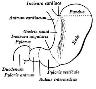
Anatomical consideration of FB impaction in esophagus.

|
What percentage of children require intervention?
Most of the fb pass spontaneously.
10- 20% require endoscopic intervention.
Less than 1% require surgical intervention.
How do the patients present ?
Smaller children are unable to provide history
of ingestion thus many of the cases may be asymptomatic and presentation may
get delayed (30% are without symptoms)
Parents may provide history of witnessing the
event
Older children and adults are able to provide
history
What are the common symptoms?
Dysphagia and odynophagia are the most common
symptoms
Other include—drooling ,refusal to eat,
globus(sensation sthg sticking in the throat)
Retrosternal chest pain
Respiratory symptoms like stridor, choking,
dyspnea, wheezing--- indicating compression of trachea by FB in
esophagus
In case of perforation—neck swelling,pneumomediastinum, crepitus in neck
Features of shock- tachycardia, cold
extremities, poor pulse volume and hypotension in cases of perforation.
Long standing esophageal FB can present with
weight loss
recurrent aspiration pneumonia
esophageal strictures
tracheo esophageal fistula
erosion into aorta
History is
important for diagnosis and the nature of object ingested also provides better
understanding of location of impaction
How can physical examination help ?
For supracricoid FB-- adequate visualization of the oral cavity, nasal passages,
pharynx and larynx is crucial for ruling out an FB in the upper aerodigestive
tract.
Tongue depressor, transnasal flexible endoscopy, indirect laryngeal mirror, Mackintosh laryngoscope and flexible pharyngolaryngoscopy may subsequently be utilized.
Tongue depressor, transnasal flexible endoscopy, indirect laryngeal mirror, Mackintosh laryngoscope and flexible pharyngolaryngoscopy may subsequently be utilized.
For
infracricoid follow up and other diagnostic and therapeutic modalities are required.
How do we investigate ?
X-rays
CT/MRI
USG
CT/MRI
USG
Fluoroscopic study
What is the use of X ray?
Both AP and lateral view X rays are necessaryX ray of chest and abdomen is necessary
How to differentiate between tracheal and esophageal?
Flat objects in esophagus orient in coronal
plane and appear circular in AP view .
In trachea they tend to orient in sagittal plane and appear circular in lateral view.
These tendencies aren’t universal thus X rays
are to be carefully evaluated.
Lateral view also helps in identifying if
multiple FBs are stacked over one another or not.

Are X rays useful in case of radiolucent FB?
It is better to do X ray even in cases of radiolucent FB as
they can provide some information like
· Air fluid level in esophagus.
· Free air s/o perforation.
X rays of Toys
with multiple components should be carefully interpreted as they contain both
radioopaque and radiolucent components.
What is the use Computed tomography?
· Symptomatic patients in whom FB localization is difficult.
· In cases of large FB i.e. more than 5 cm in
length and 3 cm in width or in cases of sharp objects
· Ct with 3 D reconstruction is useful.
Indications of MRI
MRI can be used for radiolucent FB but should be
avoided in cases of metallic foreign body
USG- not used often but can be useful in the presence of
expert radiologist .
What is the use of fluoroscopic UGI study?
It can be used for identification of location of
radiolucent FB.
· Water soluble contrast media is used
· Avoid using barium as it can obscure the view in
subsequent endoscopy.
What are the indications of urgent removal ?
Symptoms of respiratory compromise
· Evidence of near complete obstruction of
esophagus
· Ingestion of sharp or long(>5cm) or
superadsorbent objects
· Button batteries in esophagus
· Ingestion of high powered magnet or magnets
· Sign and symptoms of esophageal inflammation
ANY OBJECTS IN ESOPHAGUS FOR > 24 HOURS SHOULD BE REMOVED
IRRESPECTIVE OF TYPE OF FB
Role of proteolytic enzymes like papain or glucagon ?
Not
recommended
What is the role of proteolytic enzyme and why isn’t it recommended?
Particularly useful for food bolus/meat as it
can cause digestion and breakdown of bolus
· Reports of esophageal erosion, perforation ,
aspiration pneumonia and hypernatremia have prevented its use.
What is the role of glucagon?
It relaxes the LES and helps in passage of food
bolus.
· 2 small RCTS and a large case series have found
that glucagon no more effective than placebo in FB
· It is useful in people with history of solid
food dysphagia.
SPECIFIC MANAGEMENT FOR SPECIFIC FB.
COINS
·
In asymptomatic patients expectant management
for first 24 hrs as 20-30 % cases coin spontaneously passes to stomach.
·
In Asymptomatic cases if coin persists after 24
hrs in esophagus or if time of ingestion isn’t known removal is indicated
·
In symptomatic patients removal of FB is done as
soon as possible.
What to do for coins in stomach?
If asymptomatic—follow up with weekly X rays as
most of them pass but if the coin is in the stomach even after 4 weeks
endoscopic removal.
· Symptomatic cases require immediate removal.
How do we remove coin?
· Rigid endoscopy for proximal FB and flexible for
distal
· Several case series have shown use of bougienage
to push the coin into the stomach for selected patients as effective
alternative.(TABLE 2)
· Some radiologists use Foleys catheter for removal
during fluoroscopy with patient in prone. (TABLE 1)
SHARP OBJECTS
·
Immediate endoscopic removal using retrivel
forceps
·
Orient the object with the sharp end trailing
behind or use a protective hood on the end of the endoscope.
·
Sharp objects in stomach and duodenum should be
removed immediately
·
Some Sharp objects (closed safety pin) can be
managed like coins
·
Sharp objects in small intestine can be followed
up daily with radiographs in asymptomatic cases and surgical intervention is warranted if no signs of elimination is found even after 3 days
·
Symptomatic cases with sharp objects in
intestine warrant immediate intervention.
LONGER OBJECTS
·
Objects longer than 5 cm cannot pass the stomach
and thus should be removed.
·
Also longer objects even if they pass stomach can
get impacted in the ileocaecal jxn hence removal is ideal
FOOD IMPACTION
·
Same as coin
LEAD CONTAINING OBJECTS
·
As soon as possible
·
PPI/H2 blockers can be used as they prevent
dissolution of lead


**
Button battery and magnet is discussed in separate blog.


Notes are good. Easy to understand. Till now the best one to know the key points. Also shows how to study, what should be known in the given topic. All the best.would love to see more topics as well.
ReplyDeleteThe color selection should be changed in diagram.