Posts

Congenital Hypertrophic Pyloric Stenosis

Image
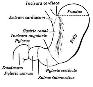
What are the parts of pylorus? The incisura angularis divides the stomach into a body to the left and a pyloric portion to the right. The sulcus intermedius further divides the pyloric portion of the stomach: the pyloric vestibule to the left, denoted by an outward convexity of the greater curvature. The pyloric antrum or pyloric canal to the right The pyloric antrum is 2.5 cm and terminates in pyloric orifice into duodenum.   What happens to the normal anatomical structural in CHPS? In infants with IHPS, the pyloric ring is no longer identifiable as a clearly definable separation between the normally distensible pyloric antrum and the duodenal cap. Instead, a channel of variable length (1.5–2.0 cm) corresponding to the pyloric canal separates the normally distensible portion of the antrum from the duodenal cap. ·          Infantile hypertrophic pyloric stenosis  was first fully described by  Harald Hirschsprung  in 1888. ·          IHPS VS CHPS - ????? IHPS is would b

Congenital Hypothyroidism: Screening and Management (AAP 2022 Recommendations)

Image
  Hypothyroidism Newborn screening (NBS) for CH followed by prompt initiation of levothyroxine (L-T4) therapy can prevent severe intellectual disability, psychomotor dysfunction, and impaired growth. The influence of severity at diagnosis, timing of treatment initiation, and L-T4 dose on longterm outcomes remains a matter of debate. Although the majority of treated patients achieve normal cognitive outcomes, the influence of L-T4 therapy on neurologic development is less certain, and some studies show persistent deficits in treated patients compared with euthyroid healthy controls. If a permanent form of CH has not been established, L-T4 treatment is maintained until 3 years of age, after which thyroid function is reevaluated following a 4- to 6-week discontinuation of L-T4. Most common cause - thyroid dysgenesis, dyshormonogenesis. Less common cause - maternal antithyroid medications, TSH receptor-blocking antibodies (TRBAb) acquired through transplacental passagematernal or infant io

Febrile Seizure: Review

Image
Febrile Seizures   What is febrile seizure? Febrile seizures are seizures that are caused by a sudden spike in body temperature with fevers greater than 38C or 100.4F, with no other underlying seizure-provoking causes or diseases such as the central nervous system (CNS) infections, electrolyte abnormalities, drug withdrawal, trauma, genetic predisposition or known epilepsy. There shouldn't be any prior history of afebrile seizure as in such case the fever might be the cause of breakthrough seizure. however , there are no consensus regarding inclusion or exclusion of children with neurodevelopment disorder such as Autism spectrum disorder which has the predisposition to seizures. Age of presentation: As per American academy of Pediatrics: 6 months to 5 years (however cases unto 7 years of age have been diagnosed as FS) As per earlier definition ILAE - infant up to age of 1 month can be diagnosed as FS. As per NIH (National Institute of Health) children between 3 months and 5 years

DNA - Basic structure

Image
  Parts of DNA Phosphate group + sugar + Base (nitrogenous base) Difference between sugar of DNA and RNA DNA has deoxyribose sugar(1 OH group attached to sugar and RNA has ribose sugar (2 OH groups) Nucleotide and nucleoside Nucleotide = Phosphate group + Base + sugar Nucleoside = Base and sugar Types of Nitrogenous base Purines and Pyrimidines Purines consists of Pure As Gold = Adenine and Guanine Pyrimidine consists of CUT the pyramids Cytosine Uracil (only in RNA) Thymine Purine nucleosides are Adenosine and Guanosine Pyrimidine nucleosides are Cytidine, Uridine and Thymidine Purine nucleotides Adenosine monophosphate, Guanosine MP How is DNA packed inside nucleus ? In the form of chromatin which is further condensed to form chromosomes What is a nucleosome ? DNA coiled twice around an octamer of Histone composed of (H2A, H2B, H3, H4) x2 with linker H1 protein. Positive and Negative charge of Chromatin Positive charged =  Lysine  and  Arginine  of Histone which bind to negative char

Study Session

Image

Study with me

Image
  Lets study 

Treatment of Wilson disease

Image
 What is the aim of treatment of Wilson disease? Removal or copper deposited in various organ and Prevention of reaccumulating. Along with the pharmacological therapy people with Wilson disease should reduce the consumption of Copper containing food items.  For Clinical features and diagnosis of Wilson Disease  First introduced in 1956 AD and still the standard of treatment. How does it act? The sulfhydryl group in d-penicillamine chelates copper removes the copper from the tissue and out of the body via urinary excretion. It can also induce the endogenous hepatic metallothionein, a cytosolic metal-binding protein, which sequesters copper and thereby limiting damage to the liver.   How penicillamine is absorbed? Its absorption is decreased by as much as 50 percent when taken with food. Thus, it should be taken 1 hour before or 2 hours after meal. What is the dose of D-penicillamine? The drug is introduced at a dose of 250 to 500 mg/day and then increased by 250 mg every 4 t