Posts

Treatment of Wilson disease

Image
 What is the aim of treatment of Wilson disease? Removal or copper deposited in various organ and Prevention of reaccumulating. Along with the pharmacological therapy people with Wilson disease should reduce the consumption of Copper containing food items.  For Clinical features and diagnosis of Wilson Disease  First introduced in 1956 AD and still the standard of treatment. How does it act? The sulfhydryl group in d-penicillamine chelates copper removes the copper from the tissue and out of the body via urinary excretion. It can also induce the endogenous hepatic metallothionein, a cytosolic metal-binding protein, which sequesters copper and thereby limiting damage to the liver.   How penicillamine is absorbed? Its absorption is decreased by as much as 50 percent when taken with food. Thus, it should be taken 1 hour before or 2 hours after meal. What is the dose of D-penicillamine? The drug is introduced at a dose of 250 to 500 mg/day and then increased ...

WILSON DISEASE

Image
  Aka Hepatolenticular degeneration  Autosomal Recessive inheritance  What is the defect in Wilson disease? Basic defect in hepatocellular copper transport, leading to the accumulation of copper in the liver and other tissues, including the brain.  ATP7B is a copper-transporting intracellular protein affected in Wilson disease. ATP7B is expressed mainly in hepatocytes.  This defect leads to reduced incorporation of copper into apo ceruloplasmin and decreased transport of copper from the liver into bile, leading to copper excess in tissue.  Although the ceruloplasmin levels are diagnostic in Wilson disease it does not play any significant role in the disease pathogenesis. Note: Hereditary aceruloplasminemia is a disease condition characterized by iron deposition in tissues and not copper  How copper deposition occurs in other tissues? Once the hepatocytes are filled with copper this leads to injury thus liberating the copper in circulation which leads t...

VACTERL/VATER ASSOCIATION

Image
  Why not syndrome and why association? VATER or VACTERL is not a syndrome as there is a no single pathological cause responsible for the common incidence. Components of VACTERL are V - Vertebral anomalies occur in 80 t0 85% cases - hemivertebrae or hypoplastic vertebrae A - Anal atresia or imperforate anus occur in 50-55% cases C - cardiac malformations occur in 75% cases VSD, ASD and ToF are common TE - tracheoesophageal fistula with esophageal atresia occur in 70% cases Renal anomalies renal anomalies occur in 50% cases like agenesis or dysgenesis, single umbilical artery can also occur Limb anomalies Radial anomalies like radial aplasia, hypoplastic thumbs can occur. Syndactyly, polydactyly can also occur.

Gitelman

Image
 Autosomal Recessive condition  More common than Bartter syndrome  Mimics chronic thiazide therapy side effects  It affects Na-Cl channel of Distal convoluted tubule Characterized by  Polyuria Metabolic alkalosis Hypokalemia Hyponatremia Hypomagnesaemia Low blood pressure  Pathophysiology of these features are similar to Bartter syndrome  For Bartter notes  How is Gitelman syndrome different from Bartter syndrome? Bartter syndrome show poor response to a loop diuretic, while patients with Gitelman syndrome have poor response to a thiazide diuretic. Concentrating ability is impaired in Bartter syndrome but is relatively preserved in Gitelman syndrome since LoH is more concerned with generating concentration gradient in kidney. Urinary calcium excretion is typically reduced in patients with Gitelman syndrome as it is with a thiazide diuretic but high in Bartter syndrome. Treatment is similar to Bartter syndrome.  

Bartter Syndrome

Image
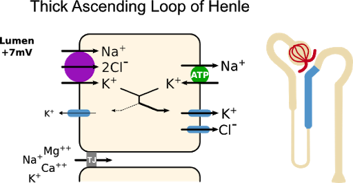
 Autosomal Recessive disorder. Characterized by defect in the Na-K-Cl channel in the thick ascending limb of loop of Henle  This leads to loss of Na - K - Cl into the urine.  Hypovolemia occurs which causes secondary hyperaldosteronism by activation of Renin-Angiotensinogen - Aldosterone mechanism. This Na-K-Cl channel is responsible for creating the medullary concentration gradient that is required for excretion of maximally concentrated urine in the presence of ADH.   The impaired concentrating ability and polyuria in Bartter syndrome are mainly due to the impaired sodium transport in the loop of Henle.  High prostaglandin levels and chronic hypokalemia may also contribute.   Why metabolic alkalosis and hypokalemia in Bartter? Volume depletion leads to secondary hyperaldosteronism; this, along with increased distal flow and delivery of sodium, results in increased urinary potassium losses and hydrogen ion secretion. Why hypocalcemia and hypomagnesaem...

Lissencephaly

Image
  Also known as agyria Some 65% are associated with mutations in the LIS1 gene. Smooth brain with no or little gyrus in the cerebral cortex It is due to incomplete or complete failure of neuronal migration during the 12th to 24th postmentrual weeks Microcephaly, ventriculomegaly, wide Sylvian fissures, complete or partial agenesis of the corpus callosum 2 types of Lissencephaly A. Type 1 is associated with facial dysmorphism sometimes with deletion of 17p chromosome known as Miller Dicker syndrome B. Type 2 is associated with hydrocephaly and dysgenesis of cerebellum - Walkar-warburg syndrome Prenatal diagnosis of lissencephaly Transvaginal ultrasound - imaging of the surface of the cerebral hemispheres and should facilitate in-utero diagnosis. Prenatal diagnosis of lissencephaly probably cannot be made reliably until 26 to 28 postmenstrual weeks, when the normal gyri and sulci become well defined. Up to this time, the normal fetal brain has a smooth appearance

Moebius syndrome

Image
Congenital unilateral or bilateral paralysis of VII and VI nerve  Hypoplasia of  nerve nuclei in the brainstem results in the condition Other cranial nerves like III, IV, V,VIII may also be involved  Exact cause is unknown.  May have - foot abnormalities like club foot  Mental retardation may be present